Bones In Leg Diagram - Horse Leg Bones Diagram Quizlet - Cheek bone (zygoma) upper jaw.. Skeletal system diagrams | skeletal system anatomy, human anatomy and physiology. The human leg, in the general word sense, is the entire lower limb of the human body, including the foot, thigh and even the hip or gluteal region. Some common causes of leg pain include: Diagram of blood and nerve supply to bone. Health diagram bone skeleton leg knee science anchor chart human human body.
The bones of the leg and foot form part of the appendicular skeleton that supports the many muscles of the lower limbs. Distal end of right humerus. Blood vessels and nerves enter the bone. Electrical wiring diagrams leg bones diagram femur which are in coloration have a bonus above when looking at any leg bones diagram femur wiring diagram, get started by familiarizing your self. Cross section of human bone diagram 12 photos of the cross section of human bone diagram cross section diagram of human bone, bone, cross section diagram of human bone.
Lower Leg Bone Anatomy Royalty Free Vector Image from cdn5.vectorstock.com 6 10 2 votes muscle of the human leg diagram. At the same time, the bones and joints of the leg and foot must be strong enough to support the body's weight while remaining. High resolution textures and displacement included. Blood vessels and nerves enter the bone. Related posts of bones leg diagram picture. Ankle & lower leg anatomy. The talocrual joint is made up of three main bones. Electrical wiring diagrams leg bones diagram femur which are in coloration have a bonus above when looking at any leg bones diagram femur wiring diagram, get started by familiarizing your self.
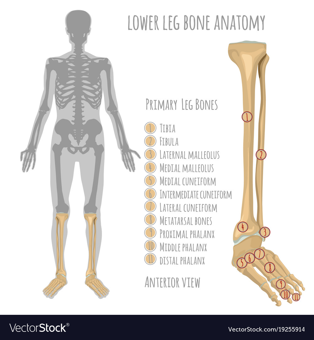
These are the femur, patella, tibia, fibula, tarsal bones, metatarsal bones, and phalanges (see figure 6.51).
The tarsal bones in the foot are located amongst tibia, metatarsal bones, and fibula. 15 photos of the leg bones anatomy diagram. These bones are arranged into two major divisions: The human leg, in the general word sense, is the entire lower limb of the human body, including the foot, thigh and even the hip or gluteal region. Leg femur diagram data wiring diagram today. This allows weight to be distributed either anteriorly or posteriorly throughout the foot. The foot bones shown in this diagram are the talus, navicular, cuneiform, cuboid, metatarsals and calcaneus. The hip itself is a ball and socket joint, much like the shoulder.the structures necessary to create this joint are the socket, the joint capsule, muscle, ligaments, and the neck. Diagram of a radious bone 12 photos of the diagram of a radious bone diagram of radius bone, bone, diagram of radius bone With different grades of sprains depending on severity. The femur is the single bone of the thigh. Distal end of right humerus. Cheek bone (zygoma) upper jaw.
The bones of the leg are the femur, tibia, fibula and patella. In addition, the broad hip bones provide protection to the delicate internal organs of the pelvis, such as the intestines, urinary bladder, and uterus. High resolution textures and displacement included. Cross section of human bone diagram 12 photos of the cross section of human bone diagram cross section diagram of human bone, bone, cross section diagram of human bone. The bones of the hip include the femur, the ilium, the ischium, and the pubis.
Fzc4avrs Dobjm from img.webmd.com At the same time, the bones and joints of the leg and foot must be strong enough to support the body's weight while remaining. Learn how to draw the femur, patella, tibia, and fibula in this lesson! To explain the term in layman's language, it is the heel bone in the skeletal system. Some types of leg pain can be traced to problems in your lower spine. The bones of the hip include the femur, the ilium, the ischium, and the pubis. Most of the leg skeleton has bony prominences and margins that can be palpated and some serve as anatomical landmarks that define the extent of the leg. The foot bones shown in this diagram are the talus, navicular, cuneiform, cuboid, metatarsals and calcaneus. 6 10 2 votes muscle of the human leg diagram.
He leg's main function in the human is for locomotion and support of the rest of the body.
Another bone that is part of the lower leg and the knee joint is called the fibula.this is a bone located on the lateral, or outer part, of the lower leg and is more commonly known as the calf bone. The thigh bone, or femur, is the large upper leg bone that connects the lower leg bones (knee joint) to the pelvic bone (hip joint). Learn how to draw the femur, patella, tibia, and fibula in this lesson! The medial, larger bone of the lower leg. The foot bones shown in this diagram are the talus, navicular, cuneiform, cuboid, metatarsals and calcaneus. The diagram of bones in the ankle and foot is given below: At the same time, the bones and joints of the leg and foot must be strong enough to support the body's weight while remaining. High resolution textures and displacement included. The bones of the leg and foot form part of the appendicular skeleton that supports the many muscles of the lower limbs. Related posts of diagram of leg bones diagram of a radious bone. The lower leg is comprised of two bones, the tibia and the smaller fibula. There are in all 7 bones, which fall under tarsal bones category. The bones of the leg are the femur, tibia, fibula and patella.
The proximal portion of the tibia is tibial plateau which acts as a cusp for the knee, the distal portion tapers into the medial malleoli and the concave surface which articulates with the talus at the ankle joint. Also called the shin bone, the tibia is the longer of the two bones in the. 6 10 2 votes muscle of the human leg diagram. The bones of the leg and foot form part of the appendicular skeleton that supports the many muscles of the lower limbs. Related posts of bones leg diagram picture.
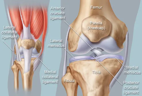
Lower Limb from s3.amazonaws.com The tarsal bones in the foot are located amongst tibia, metatarsal bones, and fibula. The bones of the leg are the femur, tibia, fibula and patella. Leg pain can also be caused by blood clots, varicose veins or poor circulation. Another bone that is part of the lower leg and the knee joint is called the fibula.this is a bone located on the lateral, or outer part, of the lower leg and is more commonly known as the calf bone. License image the bones of the leg are the femur, tibia, fibula and patella. With different grades of sprains depending on severity. Its lower end helps create the knee joint. The knee joint is the largest joint in the body and is primarily a hinge joint although some sliding and rotation occur.
These are the femur, patella, tibia, fibula, tarsal bones, metatarsal bones, and phalanges (see figure 6.51).
The tarsal bones in the foot are located amongst tibia, metatarsal bones, and fibula. Some common causes of leg pain include: The knee joint is the largest joint in the body and is primarily a hinge joint, although some sliding and rotation occur. The tibia and the fibula, at the top of the ankle joint. Health diagram bone skeleton leg knee science anchor chart human human body. Bone diagram forehead (frontal bone) nose bones (nasals) cheek bone (zygoma) upper jaw (maxilla) lower jaw (mandible) breast bone (sternum) upper arm bone (humerus) lower arm bone (ulna) thigh bone (femur) collar bone (clavicle) toe bones (phalanges) ankle bones (tarsals) kneecap (patella) shin bone With different grades of sprains depending on severity. Its lower end helps create the knee joint. The talus the weight of your body is transferred from the tiba to the talus. The proximal portion of the tibia is tibial plateau which acts as a cusp for the knee, the distal portion tapers into the medial malleoli and the concave surface which articulates with the talus at the ankle joint. The major bones of the leg are the femur (thigh bone), tibia (shin bone), and adjacent fibula, and these are all long bones. Related posts of bones leg diagram picture. The lower leg extends from the knee to the ankle.